Focus on mobile health: Innovative microscope enables early cancer diagnoses
January / February 2020 | Volume 19, Number 1
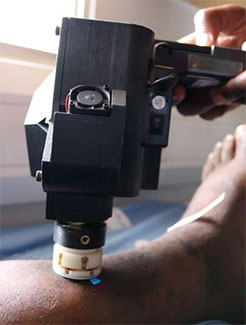
Photo courtesy of Makerere University
A smartphone confocal microscope, developed
by a Fogarty grantee, shows promise in providing
quick, inexpensive diagnoses of Kaposi’s
sarcoma lesions.
By Susan Scutti
Smartphone microscopes are showing great potential for enabling instant and affordable diagnoses of certain cancers in low-resource settings.
Fogarty grantee Dr. Dongkyun Kang has developed a new method of deploying confocal microscopy technology that provides instant images on a low-cost mobile device with support from Fogarty's
Mobile Health: Technology and Outcomes in Low and Middle Income Countries program. In a pilot study in Uganda, the device was used to examine participants' skin lesions and samples to look for
Kaposi's sarcoma (KS). Promising results led Kang to refine the prototype to improve image quality. He also has a
second Fogarty grant to develop the confocal technology to produce and study an inexpensive smartphone endoscope to screen for cervical cancer.
“We're streamlining the process to enable onsite, real-time, single-visit diagnoses," says Kang, a biomedical engineering professor at the University of Arizona. “Now the patient doesn't have to come back to the rural clinic or hospital. We can catch them when they are there."
Although confocal microscope technology has been around for several decades, it's been cost prohibitive for use in low- and middle-income countries (LMICs). The materials for
Kang's devices cost only about $4,200, as compared to nearly $100,000 for a commercial confocal microscope, he says. In addition, his latest prototype weighs only about 2 pounds, so it is portable and easy to hold with a single hand.
Improving access to and speed of diagnoses for cancer could have an enormous health impact. KS is the most common HIV-associated cancer among patients who live in sub-Saharan Africa. Survival rates are poor: In some African countries, only 30% of patients live even three years following diagnosis. The cervical cancer picture is equally bleak, with more than 80% of the world's diagnoses occurring in LMICs. In East Africa, the mortality rate is 27.6 per 100,000.
“The standard way of diagnosing any cancer is to take a little bit of tissue out of a patient and then process it as a thin section slide that a pathologist takes a look at under the microscope," explains Kang. With his smartphone device, he is bringing the microscope to the tissue rather than the tissue to the microscope, saving the usual four-to-six weeks' processing time. Kang's device joins a smartphone with an optical attachment in a custom-built, optomechanical holder. It can generate two-dimensional pictures of suspected KS lesions by using light plus a prism-like component - diffraction grating - and the image sensor in a smartphone.
His pilot study of the technology conducted at Makerere University in Kampala revealed issues with poor image quality, Kang confides. Based on these results, he refined the prototype, which now uses near-infrared (NIR) light and a new optics design.
The NIR version, which can be used for biopsies as well as on living patients, “reduces the speckle noise significantly and will have better imaging performance," according to Kang, who is planning to test the new iteration of the device soon.
His newest invention, for diagnosing cervical cancer, filters light through a confocal module and sends this light through a fiber bundle within an endoscopic catheter to the cervix. The returning light waves are then bent and focused to generate an image on the smartphone camera.
Kang’s lab has already begun to explore the use of the smartphone microscope to diagnose cervical precancers with co-investigator Dr. Miriam Nakalembe of Makerere, while the team expects to begin field tests in Kampala later this year. Nakalembe and others working on the project have received additional support from the NIH’s National Cancer Institute (NCI) and the National Institute of Allergy and Infectious Disease (NIAID).
If his smartphone confocal device is successful, Kang envisions a simplified, cost-effective “screen and treat” approach for cervical cancer: First, women will undergo the usual vinegar test; those who test negative will be scheduled for a future follow-up, while those who test positive will be examined with the confocal endoscope and, if abnormal cells are seen, these women can be treated immediately.
The diagnostic process for suspected Kaposi's sarcoma lesions also might be streamlined, says Kang: “We can take a look at the tissue on the spot - no biopsy needed, no return visit needed - so a clinician can decide whether to treat or monitor immediately.”
The endgame of his research is to develop an app powered by artificial intelligence to analyze images and either guide or provide a diagnosis. His work in Uganda has already established an mHealth knowledge base and a network of collaborating researchers in the country, which Kang believes will pave the way for
future projects and innovations across Africa.
Not only will the smartphone confocal microscope make a difference in LMICs, it will also benefit Americans, says Kang, who has begun work with an Arizona clinic that provides health care to people who lack medical insurance. “The whole goal is to make an impact in the U.S. as well."
More Information
- More about Dr. Dongkyun Kang's grants, both supported by Fogarty's
Mobile Health: Technology and Outcomes in Low and Middle Income Countries program:
-
Kaposi's sarcoma treatment information and resources from NIH's National Cancer Institute (NCI)
-
Smartphone confocal microscopy for imaging cellular structures in human skin in vivo
Biomedical Optics Express via PubMed, March 26, 2018 -
Low-cost, high-speed near infrared reflectance confocal microscope
Biomedical Optics Express via PubMed, June 21, 2019 -
Smartphone epifluorescence microscopy for cellular imaging of fresh tissue in low-resource settings
Biomedical Optics Express via PubMed, December 6, 2019 -
Dongkyun “DK” Kang, Ph.D.
University of Arizona Wyant College of Optical Sciences -
Mobile health news, resources and funding for global health researchers from Fogarty
To view Adobe PDF files,
download current, free accessible plug-ins from Adobe's website.